

Veterinary Radiography and Ultrasonography
Many diseases and medical conditions cause similar symptoms in pets. As a result, obtaining information beyond blood or urine testing is sometimes necessary for a veterinarian at Signature Veterinary Services to make an accurate diagnosis and administer appropriate treatment(s). Radiography (x-rays) and ultrasound are imaging modalities that enable veterinarians to view a pet’s internal organs and bone structure and thereby facilitate making a diagnosis and instituting therapy.
What are Radiography and Ultrasonography?
Radiography and ultrasonography are safe, non-invasive tools that veterinarians use to diagnose a variety of diseases and conditions in both cats and dogs. They are complementary diagnostic modalities; it is not uncommon for both to be utilized to obtain a diagnosis.
Veterinary Radiography
Radiographs, commonly known as x-rays, are black and white two-dimensional images. Radiation is passed through a particular structure (i.e., limb) or region (i.e., chest) to generate these pictures. The denser the tissue/structure, the whiter the image; consequently, bones are whiter and lungs are blacker. We use radiographs to survey organ size and shape, bone and joint structure, fluid accumulation and also to screen for foreign objects.
Radiographs used to be exclusively captured on film, but with technological advances, digital radiography has become mainstream in veterinary hospitals and has simplified image storage and sharing. Radiography is generally the most common and readily available diagnostic tool in a traditional veterinary hospital setting. At Signature Veterinary Services, we partner with a mobile x-ray service to provide this diagnostic modality at the home.
Veterinary Ultrasonography
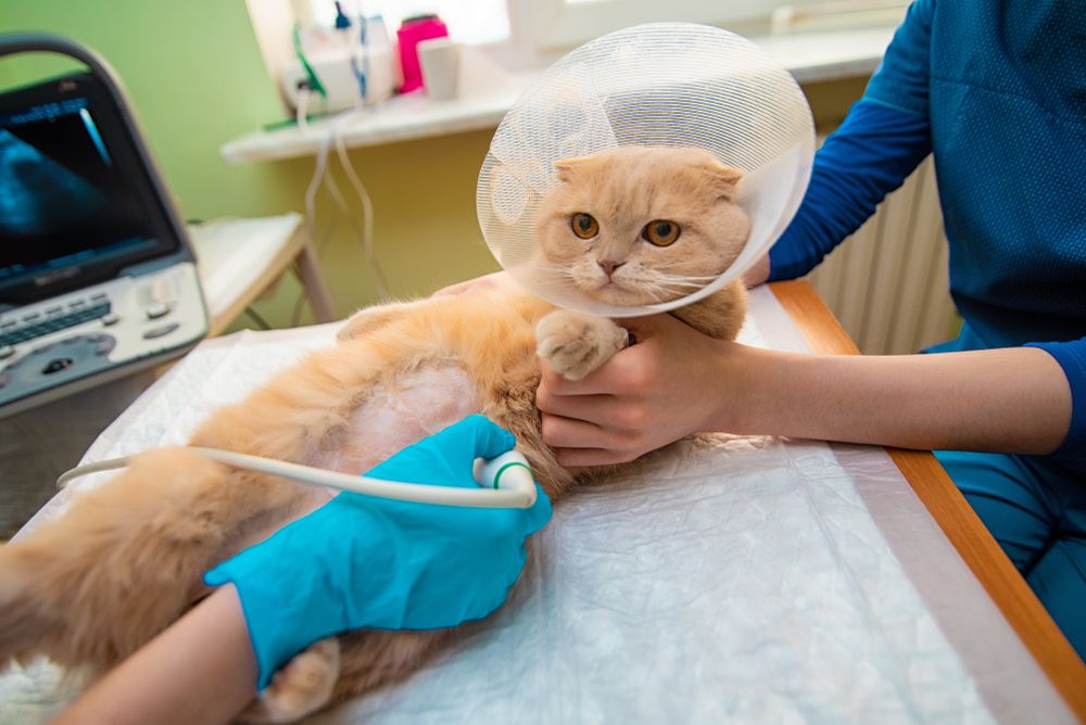
Ultrasound uses high-frequency sound waves to generate images. The waves move out from a wand (a probe), that is in contact with an area or structure of interest, and are absorbed, transmitted, and/or reflected to produce an image. Real-time and still images are displayed and captured. Ultrasound is useful for evaluating organs, fluid, and soft tissue structures and is routinely used to collect diagnostic urine samples. A specific ultrasound called an echocardiogram is the gold standard for the assessment of heart structure and function.
Ultrasound is a painless, well-tolerated procedure. While an ultrasound exam does not require anesthesia or even sedation in most cases, it does necessitate that the pet’s fur is shaved from the evaluation area. Shaving the fur enables better contact between the probe and the area of interest and thus provides better image quality.
At Signature Veterinary Services, we have a portable ultrasound machine that we use for exams on our mobile clinic or inside the home. We also have access to specialists to whom we can refer when needed. For echocardiograms, we partner with a mobile service and cardiologist to provide a comprehensive work-up at the home.
What to Expect During Your Pet's Diagnostic Appointment
Since these are non-invasive, painless procedures, many pets do not require sedation for their appointments. If a pet is anxious or has a difficult time remaining still for our veterinarian to produce or obtain a clear image, sedation might be necessary. We always provide the option of sending radiograph and ultrasound images to a radiologist for review.
Learn More About Our Mobile Veterinary Diagnostics
Signature Veterinary Services takes pride in the ability to provide our pet patients with complete care right outside their doors on our mobile clinic. We’re fully equipped to perform radiographs, ultrasounds, and echocardiograms inside our mobile clinic. For more information about diagnostic testing or to learn more about your pet’s upcoming appointment, we welcome you to contact us today.